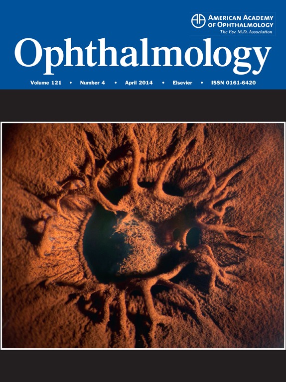
Questo studio randomizzato e controllato riporta gli esiti favorevoli del trattamento con cross-linking nella gestione del cheratocono progressivo.
La metodica si conferma efficace e affidabile a distanza di tre anni, in particolare confrontando i risultati con i cambiamenti avvenuti nello stesso tempo negli occhi non trattati.
Christine Wittig-Silva, MD, Elsie Chan, FRANZCO, Fakir M.A. Islam, PhD, Tony Wu, BSc, Orth&OphSc, Mark Whiting, FRANZCO, Grant R. Snibson, FRANZCO
Ophthalmology
Volume 121, Issue 4 , Pages 812-821, April 2014
Presented at: American Society of Cataract and Refractive Surgery Symposium on Cataract, IOL and Refractive Surgery, April 2013, San Francisco, California; and European Society of Cataract and Refractive Surgeons Congress, October 2013, Amsterdam, The Netherlands.
To report the refractive, topographic, and clinical outcomes 3 years after corneal collagen cross-linking (CXL) in eyes with progressive keratoconus.
Participants One hundred eyes with progressive keratoconus were randomized into the CXL treatment or control groups.
Methods Cross-linking was performed by instilling riboflavin 0.1% solution containing 20% dextran for 15 minutes before and during the 30 minutes of ultraviolet A irradiation (3 mW/cm2). Follow-up examinations were arranged at 3, 6, 12, 24, and 36 months.
Main Outcome Measures The primary outcome measure was the maximum simulated keratometry value (Kmax). Other outcome measures were uncorrected visual acuity (UCVA; measured in logarithm of the minimum angle of resolution [logMAR] units), best spectacle-corrected visual acuity (BSCVA; measured in logMAR units), sphere and cylinder on subjective refraction, spherical equivalent, minimum simulated keratometry value, corneal thickness at the thinnest point, endothelial cell density, and intraocular pressure.
Results The results from 48 control and 46 treated eyes are reported. In control eyes, Kmax increased by a mean of 1.20±0.28 diopters (D), 1.70±0.36 D, and 1.75±0.38 D at 12, 24, and 36 months, respectively (all P < 0.001). In treated eyes, Kmax flattened by −0.72±0.15 D, −0.96±0.16 D, and −1.03±0.19 D at 12, 24, and 36 months, respectively (all P < 0.001). The mean change in UCVA in the control group was +0.10±0.04 logMAR (P = 0.034) at 36 months. In the treatment group, both UCVA (−0.15±0.06 logMAR; P = 0.009) and BSCVA (−0.09±0.03 logMAR; P = 0.006) improved at 36 months. There was a significant reduction in corneal thickness measured using computerized videokeratography in both groups at 36 months (control group: −17.01±3.63 μm, P < 0.001; treatment group: −19.52±5.06 μm, P < 0.001) that was not observed in the treatment group using the manual pachymeter (treatment group: +5.86±4.30 μm, P = 0.181). The manifest cylinder increased by 1.17±0.49 D (P = 0.020) in the control group at 36 months. There were 2 eyes with minor complications that did not affect the final visual acuity.
Conclusions At 36 months, there was a sustained improvement in Kmax, UCVA, and BSCVA after CXL, whereas eyes in the control group demonstrated further progression.strong 0.001; treatment group: −19.52±5.06 μm, P 0.001; treatment group: −19.52±5.06 μm, P

